
Indian Ringspot
HISTORY, DISTRIBUTION AND IMPORTANCE
Described by Ahlawat (1989) as a disease with ringspot symptoms mainly affecting Kinnow mandarin. Thought at the time to be related to psorosis A, but there were no bark-scaling symptoms, and the flecking, vein-clearing and ringspots persisted in mature leaves, indicating that the disease was different from psorosis. First observed in Kinnow mandarin mother trees at Punjab Agricultural University, Abohar. This material was initially imported and all the Kinnow mandarin in the country has been propagated from this source. The presence of 100% infection in Kinnow mandarin suggests that the virus might have been introduced through the imported material. Flexuous filamentous virus-like particles were found in leaf extracts of graft-inoculated Mosambi sweet orange (Byadgi et al., 1993) and the disease was further studied by Byadgi & Ahlawat (1995). The virus was mechanically inoculated to Chenopodium quinoa, producing local lesions, and to French bean Phaseolus vulgaris cv ‘Saxa’, giving systemic infection; it was purified and partially sequenced; antisera and molecular reagents were prepared (Rustici et al., 2000). Mechanical inoculation had been attempted before (Byadgi & Ahlawat, 1995) but without success because greenhouse temperatures were too high. Later the viral genome was fully sequenced, and as a result a new species, Indian citrus ringspot virus (ICRSV) in the genus, Mandarivirus, within the family Flexiviridae was proposed (Rustici et al., 2002; Adams et al., 2004; Adams et al., 2005). Virus-free stock produced by shoot-tip grafting, and indexing by ISEM, ELISA, PCR and dot-blot hybridization, is now becoming available (Hoa & Ahlawat, 2004; Hoa et al., 2004).
The disease is very widespread (up to 100%) in most Kinnow mandarin orchards in northern India, especially Punjab (Ahlawat & Pant, 2003). The incidence in sweet orange, in 1989, was 20-50% in Maharashtra, Andra Pradesh and Karnataka provinces. In the rootstocks Sohsarkar (Citrus karna) and Karna khatta, incidence was around 55%, but was only 7% in Troyer citrange (Ahlawat & Pant, 2003). Not reported outside India but could be present.
Kinnow mandarin and Mosambi sweet orange, widely grown in India, are both highly susceptible. Other varieties affected are listed under Natural Host Range, below. There has been little or no healthy planting material available, with resulting very high and widespread losses to this disease, estimated as 20-98% in different areas (Byadgi & Ahlawat, 1995; Hoa & Ahlawat, 2004).
NAME OF DISEASE AND SYNONYMS
The only established name of the disease is ‘Indian citrus ringspot’, though in the past, names such as ‘citrus ringspot’ or ‘psorosis’ were used.
SYMPTOMATOLOGY
General aspect of affected tree
Trees develop a thin canopy, and dieback symptoms leading to death after a few years.
Symptoms on trunk, limbs and shoots
There is no bark-scaling or diagnostic symptom on trunk or limbs.
Symptoms on leaves
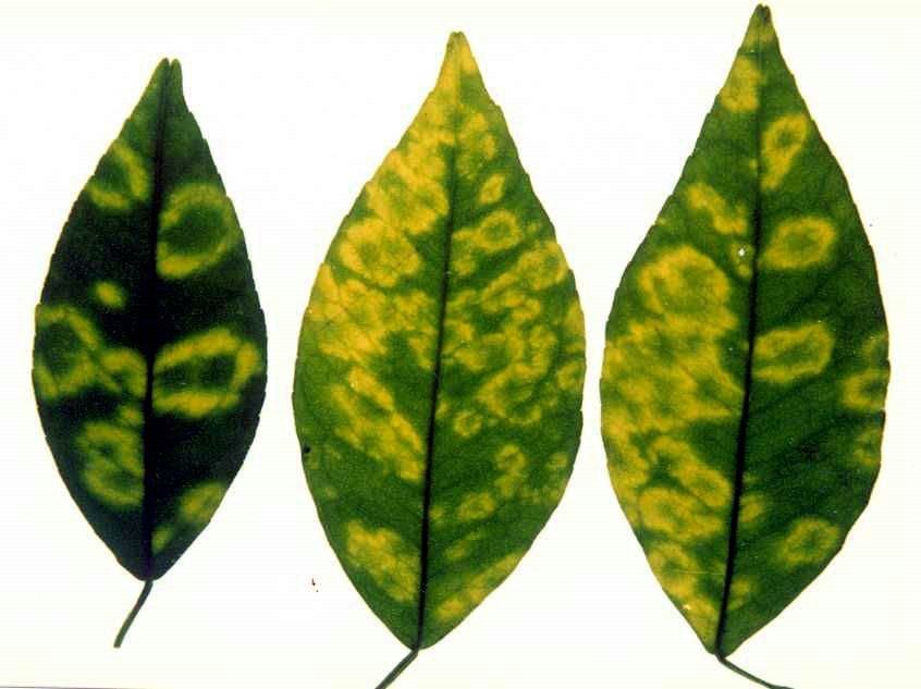
On leaves of Mosambi sweet orange, vein-clearing, vein-banding and flecking develop in young leaves but then persist in mature leaves. Ringspots may develop. In Kinnow mandarin, conspicuous yellow ringspots with green centers develop in mature leaves (Ir. 01); necrotic spots develop in leaves of graft-inoculated King mandarin (Hoa & Ahlawat, 2004). The ringspots on Kinnow are present from December to February when the temperature in northern India is low; for the rest of the year no symptoms are visible on field trees.
Symptoms on fruit
Fruit is reduced in quantity and quality, but without specific symptoms.
Histological and cytological symptoms
Histological symptoms not described. In thin sections of systemically infected ‘Saxa’ bean leaves, abundant virus particles were seen by EM in the cytoplasm (Ir. 02, 03) but without any specific or diagnostic cytopathology (Rustici et al, 2000).
CAUSAL AGENT: DESCRIPTION AND PROPERTIES
The disease appears to be caused by Indian citrus ringspot virus, the only current member of the genus Mandarivirus in the family Flexiviridae (Adams et al., 2004, 2005). However, purified virion preparations or infectious RNA have not yet been reintroduced into citrus, reproducing the disease. The virions, of 650 nm modal length and about 13 nm in diameter, are flexuous rods with a distinct zipper-like structure (Ir. 04, 05). There is a single coat protein of 34 kDa (325 aa) and one molecule of positive-strand ssRNA 7560 nt in length excluding the poly-A tail. The first five open reading frames somewhat resemble those of potexviruses but there is a sixth ORF as in carla- and allexiviruses. The particles are relatively robust, easily purified, and highly immunogenic.
It should be noted that, as abundantly documented but not widely known, in vitro preparations of leaves of many if not all types of healthy citrus contain filamentous structures that somewhat resemble rod-shaped virus particles and have sometimes been taken as such (Ir. 06). The virions of ICRSV and other viruses should not be confused with these. With citrus infected by ICRSV, a tubular component can also co-purify with the virions (Ir. 07) (Byadgi & Ahlawat, 1995; Byadgi et al., 1993) but its role in the disease is doubtful. Both these components are eliminated if the virus is passaged through Phaseolus vulgaris.
Hoa & Ahlawat (2004) have described several isolates of ICRSV. Analysis showed some variation near the N terminus of the coat protein but conservation in the core region. Isolates from Delhi (ICRSV-Dl), Abohar (ICRSV-Ab), Ahmedabad (ICRSV-Ah) and Pune (ICRSV-Pu) were compared with the original isolate (ICRSV-D, Byadgi et al., 1993; Rustici et al., 2000, 2002). Isolates Ab and Dl were closely related to D (98% and 99.5% aa sequence identity, respectively). Isolates Ah and Pu were similar to each other (99.6% identity) but showed only 85% and 84.5% identity, respectively, to isolate D. All the isolates were serologically similar. When graft-inoculated to a panel of citrus cultivars (Kinnow, Mosambi, Malta, Rough lemon and King mandarin) the different isolates induced various combinations of vein flecking, vein clearing, chlorotic spots, ringspots and necrotic spots, sufficient for initial identification of the isolates (Hoa & Ahlawat, 2004).
HOST RANGE
Natural
No resistant or tolerant citrus varieties have yet been identified. The commonly grown Kinnow mandarin and Mosambi sweet orange are highly susceptible, and the virus has also been detected in King mandarin, Willow leaf mandarin (the parents of Kinnow), the sweet oranges Pineapple and Satgudi, sour orange, Nagpur orange, Rangpur lime, Kagzi lime and Kagzi kalan (C. aurantifolia (Christen) Swingle) and Karna khatta (C. karna Raf.). Rough lemon and Malta sweet orange were also easily infected by graft-inoculation (Hoa & Ahlawat, 2004; Hoa et al., 2004).
Experimental
After mechanical inoculation, Chenopodium quinoa, C. amaranticolor, Glycine max (soybean, cv ‘Hodgson’) and Vigna unguiculata (cowpea, cv ‘Black’) develop only local lesions (Ir. 08); Phaseolus vulgaris (French bean, cv ‘Saxa’ and cv ‘Singtamy’) give local lesions (Ir. 09, 10) followed by vein clearing and systemic mosaic or mottle (Ir. 11, 12), the virus reaching high titer.
The following plants appear resistant: Cucumis sativus (cucumber, cv ‘Marketer’), Cucurbita pepo (zucchini, cv ‘Genovese’), Datura stramonium, Gomphrena globosa, Nicotiana benthamiana, N. clevelandii, N. glutinosa, N. megalosiphon, N. tabacum (cv ‘White Burley’), Petunia hybrida, Phaseolus coccineus (runner bean, cv ‘di Spagna’) and Pisum sativum (garden pea, cv ‘Nano’) (Hoa & Ahlawat, 2004; Rustici et al., 2000).
TRANSMISSION
Natural
No vector has been identified and the virus is not seed-transmitted in the citrus varieties tested or in Phaseolus vulgaris (Hoa et al., 2004, Rustici et al., 2000). The virus may have been present in the original Kinnow mandarin mother plants and has been widely spread by grafting and propagation. Kinnow trees are symptomless in warm weather and appear to offer good material for propagation.
Experimental
The virus can easily be transmitted by grafting and, among susceptible herbaceous hosts, by mechanical inoculation if greenhouse temperatures are not too high (see Experimental Host Range, above). The virus is transmissible between citrus plants by dodder; no transmission (persistent or non-persistent) was obtained with four aphid species tested: Aphis gossypii, A. citricola, A. craccivora and Myzus persicae; no transmission was obtained by seed or through soil (Byadgi & Ahlawat, 1995).
EPIDEMIOLOGY
Little data available. Natural spread may occur as seedling trees have been found infected (Ahlawat, personal observation) although no vector has been identified.
DIAGNOSIS
Diagnostic field symptoms
The clearest diagnostic symptoms, especially in the winter months, are the yellow rings with green centers that develop in mature leaves of Kinnow mandarin (Ir. 01) and also in Mosambi and Malta sweet orange. Ringspots disappear in summer. Other symptoms are chlorotic flecks and vein clearing or vein banding in young leaves, persisting in mature leaves. Bark-scaling does not occur.
Comparison with diseases showing similar symptoms
Symptoms, in early stages, may be confused with those of psorosis. The presence of conspicuous rings and blotches in adult leaves are reminiscent of the original ‘citrus ringspot’ disease of Wallace and Drake described in 1968, now known to have been caused by an isolate of Citrus psorosis virus. This symptom, however, is not typical of psorosis.
Biological indexing
This is not widely used, but grafting to the varieties Kinnow, Mosambi, Malta, Rough lemon and King mandarin should give a diagnostic panel of leaf symptoms including vein flecking, vein clearing, chlorotic spots, ringspots and necrotic spots, that will also distinguish between some strains of the virus (Hoa & Ahlawat, 2004).
Serological and molecular methods
Good antisera are available (Rustici et al., 2000, Hoa et al., 2004) and EM grids coated with antibody can easily trap the characteristic virus particles (ISEM), which can be clearly decorated. DAS-ELISA is also effective. RT-PCR and dot-blot hybridization protocols are available (Hoa and Ahlawat, 2004; Hoa et al., 2004) but large-scale validation of RT-PCR based detection has yet to be done.
CONTROL
Control of this disease, damaging and widespread in India and possibly elsewhere, is just beginning but should be effective. Production of virus-free material of Kinnow mandarin and Mosambi sweet orange using shoot-tip grafting has begun, and should also be applicable to rootstock varieties. Reliable means of detection have recently become available, and there appears to be no seed transmission. No natural vector is known and there are hopes that clean material planted in the field may remain virus-free. However, a vector is suspected because seedling trees have been found infected in nature (Ahlawat, personal observation). Legislation to prevent the virus being spread by grafting contaminated material has still to come into force.
NOTE ON CITRUS YELLOW VEIN CLEARING VIRUSES
Two citrus diseases reported from Pakistan (Grimaldi & Catara, 1996) and India (Alshami et al., 2003) may be caused by viruses related to ICRSV, although the symptoms are generally different from those of ICRSV. The Pakistani disease, originally found in Eureka Cascade lemons, causes strong yellow vein clearing and leaf distortion, with some ringspotting and vein necrosis in lemons but is also graft-transmissible to other citrus varieties. The Indian virus, first found in Etrog citron, causes similar symptoms and is also graft-transmissible to other citrus varieties. The two viruses can be mechanically transmitted to the same herbaceous hosts. Infected plants of both Pakistani and Indian origin contain flexuous particles indistinguishable from those of ICRSV, and the coat protein of the Indian virus is of a similar size to the ICRSV CP. Serological tests indicate that the Indian virus is not serologically related to ICRSV, but EM decoration tests on material from some Indian field trees showed up a mixed population of particles, some decorated with ICRSV antiserum and others not. The serological relationship of the Pakistani and Indian yellow vein clearing viruses has not been tested. Provisionally the two yellow vein clearing viruses from Pakistan and India have been given the same unofficial name but further investigation is needed understand their relationships.
SELECTED REFERENCES
Adams, M.J., J.F. Antoniw, M. Bar-Joseph, A.A. Brunt, T. Candresse, G.D. Foster, G.P. Martelli, R.G. Milne, C.M. Fauquet (2004). The new plant virus family Flexiviridae and assessment of molecular criteria for species demarcation. Arch. Virol., 149: 1045-1060.
Adams, M.J., G.P. Accotto, A.A. Agranovsky, M. Bar-Joseph, M., Boscia, et al.(2005). Family Flexiviridae. p. 1089-1124. in C.M.Fauquet, M.A. Mayo, J. Maniloff, U. Desselberger and L.A. Ball (eds) Virus Taxonomy, Eighth Report of the International Committee on Taxonomy of Viruses, Elsevier Academic Press, London.
Ahlawat, Y.S. (1989). Psorosis: a disease of citrus in India. Indian Phytopathology 42: 21-25.
Ahlawat, Y.S., Pant, R.P. (2003). Major virus and virus-like diseases of citrus in India, their diagnosis and management. Ann. Rev. Plant Pathol. (India) 2: 447-474.
Alshami, A.A.A., Y.S. Ahlawat, R.P. Pant (2003). A hitherto unreported yellow vein clearing disease of citrus in India and its viral etiology. Indian Phytopathology 56: 422-427.
Byadgi, A.S., Y.S. Ahlawat, (1995). A new viral ringspot disease of citrus (Citrus species) in India. Indian Journal of Agricultural Science,65: 763-770.
Byadgi, A.S., Y.S. Ahlawat, N.K. Chakraborty, A. Varma, M. Srivastava, R.G. Milne, 1993. Characterization of a filamentous virus associated with citrus ringspot in India. P. 155-162. In P. Moreno, J.V. da Graça and L.W. Timmer (eds) Proc 12th Conf. Intern. Organization Citrus Virol., Univ. California, Riverside.
Grimaldi, V., A. Catara (1996). Association of a filamentous virus with yellow vein clearing of lemon. P. 343-345. In J.V. da Graça, P. Moreno and R.K.Yokomi (eds) Proc. 13th Conf. Intern. Organization Citrus Virol., Univ. California, Riverside.
Hoa, N. V. and Y.S. Ahlawat (2004). Characterization of four isolates of Indian citrus ringspot virus. Indian Phytopathology, 57: 296-302.
Hoa, N.V., Y.S. Ahlawat, R.P. Pant (2004). Production of virus-free Kinnow mandarin and Mosambi sweet orange nucleus planting material through shoot tip grafting. Indian Phytopathology, 57: 482-487.
Rustici, G., G.P. Accotto, E. Noris, V. Masenga, E. Luisoni, R.G. Milne (2000). Indian citrus ringspot virus: a proposed new species with some affinities to potex-, carla-, fovea- and allexiviruses. Archives of Virology 145:1895-1908.
Rustici, G., R.G. Milne, G.P. Accotto (2002). Nucleotide sequence, genome organization and phylogenetic analysis of Indian citrus ringspot virus. Archives of Virology 147: 2215-2224.
Prepared (2007) by R.G. Milne,
Istituto di Virologia Vegetale CNR,
Strada delle Cacce 73, 10135 TORINO (Italy)
R.P. Pant
Plant Virology Unit, Division of Plant Pathology,
Indian Agricultural Research Institute, NEW DELHI 110012 (India)
Y.S. Ahlawat
Plant Virology Unit, Division of Plant Pathology,
Indian Agricultural Research Institute, NEW DELHI 110012 (India)
PHOTOS |
LEGENDS AND AUTHORS |
|---|---|

|
Indian ringspot symptoms in winter (seen by transmitted light) on mature leaves of Kinnow mandarin. (Delhi, India) – Y.S. Ahlawat
|

|
Thin section of leaf parenchyma cell of Phaseolus vulgaris (French bean) cv ‘Saxa’ systemically infected with Indian citrus ringspot virus. Vacuole above, chloroplasts at left, cell wall below. Note the anomalous bulge in the cytoplasm composed of proliferated membranes and virus particles. Bar = 1 micrometer. (Turin, Italy) – V. Masenga and R.G. Milne
|

|
Thin section of leaf parenchyma cell of Phaseolus vulgaris (French bean) cv ‘Saxa’ systemically infected with Indian citrus ringspot virus. Vacuole above, chloroplast at right, nucleus and mitochondrion at left, cell wall below. Note cytoplasm full of filamentous virus particles. Bar = 1 micrometer. (Turin, Italy) – V. Masenga and R.G. Milne
|

|
Purified preparation of Indian citrus ringspot virus from systemically infected Phaseolus vulgaris (French bean) cv ‘Saxa’. 0.5% uranyl acetate negative stain. Note the flexuous filamentous particles with a zipper-like structure. Some particles of the host plant protein Rubisco are seen in the background (disks about 10 nm in diameter). Bar = 100 nm. (Turin, Italy) – R.G. Milne
|

|
Purified preparation of Indian citrus ringspot virus from systemically infected Phaseolus vulgaris (French bean) cv ‘Saxa’. 0.5% uranyl acetate negative stain. Note the flexuous filamentous particles with a zipper-like structure. (Turin, Italy) – R.G. Milne
|

|
Flexible virus-like particles which are not virus, purified from healthy citrus leaves, negatively stained in 0.5% uranyl acetate. Such fibres can easily co-purify with particles of Indian citrus ringspot virus, somewhat similar in size and shape. Bar = 100 nm. (Turin, Italy) – R.G. Milne
|

|
A tubular component that can co-purify with particles of Indian citrus ringspot virus prepared from infected citrus. Uranyl acetate negative stain. Bar = 100 nm. (Delhi, India) – R.P. Pant
|

|
Local lesions in a leaf of Chenopodium quinoa following mechanical inoculation with a buffered crude extract of Indian citrus ringspot virus-infected citrus leaf. Lesions appear after about 10 days at 18-25 °C but the infection does not become systemic. (Turin, Italy) – R.G. Milne
|

|
Local lesions in a leaf of Phaseolus vulgaris (French bean) cv ‘Saxa’ after mechanical inoculation with a buffered crude extract of Indian citrus ringspot virus-infected citrus leaf. Lesions appear in about a week at 18-25 °C. (Turin, Italy) – R.G. Milne
|

|
Local lesions in a leaf of Phaseolus vulgaris (French bean) cv ‘Saxa’ after mechanical inoculation with a buffered crude extract of Indian citrus ringspot virus-infected bean leaf. These lesions are about two weeks old and are beginning to spread to the veins and induce systemic infection. (Turin, Italy) – R.G. Milne
|

|
First systemic symptoms of Indian citrus ringspot virus infection appearing in a leaf of Phaseolus vulgaris (French bean) cv ‘Saxa’ that was not directly inoculated. (Turin, Italy) – R.G. Milne
|

|
Well developed systemic symptoms (mosaic and mottle) of Indian citrus ringspot virus in Phaseolus vulgaris (French bean) cv ‘Saxa’. (Turin, Italy) – R.G. Milne
|